Molecular Pathology of Human Hemoglobin
Lecture by Max Perutz
- 1973-Jun
These captions and transcript were generated by a computer and may contain errors. If there are significant errors that should be corrected, please let us know by emailing digital@sciencehistory.org.
Transcript
00:00:01 ...director of the Medical Research Council Unit for Molecular Biology at Cambridge, England,
00:00:08 past chairman of the European Molecular Biology Organization,
00:00:14 and he holds honorary degrees of Doctor of Philosophy from the University of Vienna and the University of Edinburgh.
00:00:22 Dr. Perutz and I first met when we were both students at the University of Vienna,
00:00:29 where we shared an interest in chemistry, mountaineering, and skiing.
00:00:36 We both have kept up our three interests throughout the years,
00:00:42 and yesterday we had the pleasure of showing Dr. Perutz Mount Rainier in all its glory during a hike in Mount Rainier National Park.
00:00:52 This is Dr. Perutz's first visit to Seattle,
00:00:55 and we are indeed pleased and honored that we should have him as the first Philip E. Wilcox Memorial Lecturer.
00:01:04 His lecture is entitled, The Molecular Pathology of Human Hemoglobins.
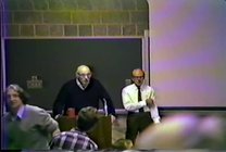
00:01:09 Ladies and gentlemen, Dr. Perutz.
00:01:12 Thank you.
00:01:34 I'm not showing this until afterwards.
00:01:39 Ladies and gentlemen, I'm very pleased to be here in Seattle,
00:01:48 and greatly honored to be asked to give a lecture in memory of a biochemist who spent much of his working life
00:01:58 trying to design methods that would help protein crystallographers to solve protein structures
00:02:06 and to get to know more about the structure and mechanism of the active sites of enzymes.
00:02:19 I want to talk today about a different kind of problem, the one of congenital diseases
00:02:29 and the contributions which protein crystallography was able to make in gaining a deeper understanding of them.
00:02:38 As you know, these are a widespread cause of human suffering,
00:02:42 and by its discovery of the genetic code and of the apparatus used for translating the base sequence of DNA into amino acid sequence,
00:02:56 molecular biology has provided us with an intellectual framework for understanding the nature of genetic diseases.
00:03:07 It appears that point mutations in the genes coding for protein chains lead to amino acid substitutions in enzymes or other proteins
00:03:19 which render these proteins inactive, so inhibiting their normal function,
00:03:30 and that this leads to the breakdown of one particular catalytic mechanism in the organism, say.
00:03:41 Now, can we go further than this?
00:03:45 The difficulty is that, apart from hemoglobin, there is yet not a single human protein of which we know both the amino acid sequence and the three-dimensional structure.
00:03:58 So, the only one where we can try and get a picture of the kind of damage that mutations do to function,
00:04:16 the only protein available so far then, is hemoglobin.
00:04:20 Now, at the same time, more variants have been discovered for this protein than for any other,
00:04:27 so that a study of the hemoglobin diseases offers us a unique opportunity for getting a better understanding of genetic effects on protein structure generally.
00:04:41 So, what do we want to do?
00:04:44 In this talk, I shall try to show you first what hemoglobin looks like, how it works,
00:04:50 and then discuss the effects of certain mutations on its structure and function.
00:04:57 Now, hemoglobin is a tetramer made up of two alpha chains, each containing about 141 amino acid residues each,
00:05:11 and two beta chains, each containing 146 residues, and four hemes.
00:05:18 And, as you know, each of these hemes is capable of combining reversibly with molecular oxygen.
00:05:25 Now, let's first look at what kind of structure one of these subunits has.
00:05:37 Tell me, have you got a pointer somewhere?
00:05:46 Oh, I'm afraid I forgot to ask.
00:05:55 Oh, here comes somebody with a device.
00:06:09 Thank you.
00:06:10 Thank you.
00:06:22 Lovely. Thank you very much.
00:06:30 Can we have these front lights off, please?
00:06:35 Now that's better.
00:06:39 This picture shows the tertiary structure of one of these globin subunits.
00:06:49 Never mind whether alpha or beta, we'll discuss that later.
00:06:53 And here is its amino end, and up there its carboxyl end.
00:06:58 Here is its amino end, and up there its carboxyl end.
00:07:02 Now, to make the journey from one end to the other, we start here,
00:07:07 then go around an alpha helix of 16 residues, turn a right-angled corner, go around a complex sort of loop,
00:07:16 then travel down one helix and up another, go through a non-helical segment,
00:07:24 and then go around this hairpin bend, and finally we arrive at the other end.
00:07:31 Now, then, the beta chain is made of eight helical segments and the same number of non-helical ones.
00:07:41 The only difference in structure between the alpha and the beta chain
00:07:45 is a deletion of two residues in this corner in the beta chain,
00:07:52 a deletion of seven residues, the C helix in the alpha chain,
00:07:58 and then finally the beta chain has one additional residue here.
00:08:02 Otherwise, these two types of structures, despite their many amino acid substitutions, are closely similar.
00:08:10 Now, this model, this drawing here, shows only the alpha-carbon positions of the chain.
00:08:18 If you look at it in three dimensions, what strikes you immediately is the exclusion of non-polar residues from the interior.
00:08:26 The interior of the chain is oily or waxy.
00:08:30 It's filled with hydrocarbons like phenylalanine, leucines, valines, tryptophanes, tyrosines,
00:08:37 and the surface is studded with charge groups, aspartic and glutamic acids, lysines, arginines, and so on,
00:08:45 which is soluble in water.
00:08:48 So, you will see the great importance of this non-polar interior later when we come to the unstable hemoglobins,
00:08:56 and you will see also how important it appears to be that all gaps in the surface are plugged, that they are closed.
00:09:04 There must not be a hole in these loops through which water can get in.
00:09:11 As soon as it does, the structure becomes unstable.
00:09:15 Now, why is this?
00:09:19 If you look at other protein structures, you'll find, generally, that the loops of the chains are strongly cross-linked
00:09:28 by hydrogen bonds between beta-plated sheets or, say, by hydrogen bonds between tyrosines and aspartates,
00:09:37 by cysteine bridges, and so forth.
00:09:40 The hemoglobins are not unique but rare in that they lack all this.
00:09:47 There are no strong bonds linking these helical regions together.
00:09:53 They just stick together by Van der Waals interactions or non-polar ones, if you like,
00:09:59 so that the hemoglobin molecules are relatively soft as protein goes.
00:10:06 You will see this has other important consequences because any misfit that a mutation produces
00:10:15 can give rise to dislocations which run right through the molecule and produce functional disturbances.
00:10:23 Now, the heme fits into this waxy mass so that its non-polar ends stick inwards and its propionic acids outwards.
00:10:36 The heme, then, is in contact with about 60 atoms of the globin,
00:10:43 and all these are Van der Waals contacts except for the one covalent link to the heme-linked histidine,
00:10:50 residue F8, which you see there.
00:10:53 And then on the other side, there's another histidine which may or may not form a hydrogen bond with the oxygen.
00:11:01 We don't really know exactly what its purpose is.
00:11:05 And then you see here the penultimate residue is a tyrosine,
00:11:10 and this tyrosine in deoxyhemoglobin, at any rate, forms a hydrogen bond with a carbonyl group of this residue there,
00:11:20 which is a valine.
00:11:21 And that, you will see, is also a very important link for stabilizing the deoxy form of this structure.
00:11:33 Now, any other points that we ought to consider while we look at this picture?
00:11:40 I don't think so.
00:11:43 So let's now look at the next one and see how two of these chains combine.
00:11:51 So here we have an alpha chain and a beta chain forming an alpha-beta dimer,
00:11:59 such as you get if you dissociate hemoglobin in strong salt solution.
00:12:05 And you see that this dimer sticks together along these helical regions here, which are the helices G.
00:12:16 The amino end of the chain is now there, the carboxyl end of the alpha chain there.
00:12:21 The amino end of the beta chain here, this carboxyl end there.
00:12:25 And here where that marker is, there's a two-fold symmetry axis,
00:12:29 which means that a rotation by 180 degrees about this vertical axis
00:12:35 would bring these two subunits into congruence with the other two that lie in front.
00:12:42 Now, between them lies a cavity, which is filled with water.
00:12:49 And between the beta chains, especially in the deoxy structure,
00:12:53 you will see there's a large hole into this 2,3-DPG fits.
00:12:59 So the next slide then shows you the complete molecule, two alpha chains, two beta chains.
00:13:13 And the form that this takes is the oxy structure,
00:13:20 where there's hardly any contact between the two alpha chains or the two beta chains,
00:13:30 but the tetramer is held together by contacts between alpha and beta here and there.
00:13:40 So there are two different kinds, alpha 1, beta 1, we call them, and alpha 1, beta 2.
00:13:48 Right. Now, what's the good of this molecule?
00:13:54 What purpose does it serve, and why does it have to be so complicated?
00:13:58 Why doesn't a single chain, say the beta chain,
00:14:03 why isn't that enough as an oxygen carrier in the red cell?
00:14:08 The answer is to be found—can we have the lights on for a moment, please?
00:14:14 The answer is to be found in the complex cooperative properties which hemoglobin has
00:14:22 and which are needed for it to fulfill its respiratory function.
00:14:27 As you know, the binding of oxygen of a hemoglobin solution is cooperative
00:14:32 in the sense that the oxygen affinity rises with increasing oxygen saturation,
00:14:39 and so it falls with decreasing oxygen saturation,
00:14:45 which seems a truism, but I say it because that is the physiologically important point.
00:14:53 It's easy to have an oxygen carrier that gets fully saturated with oxygen in the lungs,
00:15:00 where oxygen is plentiful,
00:15:03 but because the difference in partial pressure of oxygen between the lungs and the tissues
00:15:09 is only a factor of 10, it's very difficult to design an oxygen carrier
00:15:14 which will also dissociate a significant fraction of the oxygen it carries in the tissues.
00:15:22 And the cooperative properties of the hemoglobin, the sigmoid oxygen equilibrium curve,
00:15:29 allows for this.
00:15:31 So it allows a much larger fraction of the oxygen to be dissociated
00:15:36 than a hyperbolic oxygen dissociation curve such as you would get from a single chain would get.
00:15:43 So now there are several other curious properties.
00:15:48 The oxygen affinity of hemoglobin falls with falling pH.
00:15:54 It also falls with increasing concentration of various inorganic anions,
00:16:03 and especially phosphates, and especially certain organic phosphates,
00:16:11 and end of CO2.
00:16:20 So these chemically completely different agents, hydrogen ions, organic phosphates, and CO2,
00:16:31 all produce the same physiological effect.
00:16:35 They lower the oxygen affinity, and that seems like an extremely baffling property to any chemist.
00:16:42 How can three such different things produce the same effect?
00:16:49 Now, what purpose do they serve?
00:16:53 All these mechanisms help to make the discharge of oxygen yet more efficient.
00:17:02 So the presence of lactic acid and of CO2 in the tissues helps the discharge of oxygen.
00:17:11 Conversely, the uptake of hydrogen ions by hemoglobin on loss of oxygen
00:17:17 neutralizes the carbonic acid formed on combination of water and CO2,
00:17:23 and so helps to bring the CO2 into solution in the form of bicarbonate,
00:17:28 in which it can be transported back to the lungs.
00:17:31 And, of course, again, the lowering of the oxygen affinity by CO2 helps the discharge of oxygen.
00:17:39 And finally, when we come to organic phosphates, there's one specific one in human red cells,
00:17:46 2,3-diphosphoglycerate, which acts as a regulator of the oxygen affinity,
00:17:51 so that under stress, through loss of blood or going to high altitude,
00:17:59 there's a feedback mechanism producing more 2,3-diphosphoglycerate,
00:18:04 which allows for better discharge of oxygen.
00:18:11 It's the combination of these remarkable cooperative effects,
00:18:15 which have made hemoglobin such a fascinating field of study
00:18:19 for physiologists and biochemists and even physicists,
00:18:24 and these aspects, the mechanism of these, I shall be discussing in the subsequent lectures.
00:18:33 But in this talk, I just want to give you a brief outline of how it works,
00:18:38 because we need to know this in order to understand
00:18:42 how the abnormal hemoglobins interfere with the workings of hemoglobin.
00:18:48 Now, all these cooperative effects disappear if you dissociate hemoglobin into alpha-beta dimers.
00:18:57 So they're a specific property of the tetramer,
00:19:02 and not shown by either the alpha chains or the beta chains
00:19:07 or a combination of one alpha and one beta chain.
00:19:10 And that seems remarkable, that, for instance,
00:19:13 the oxygen affinity of the dimer should be independent of pH,
00:19:17 but when you assemble it into a tetramer, it becomes pH-dependent.
00:19:22 And the explanation is this, that hemoglobin exists in two different forms,
00:19:29 an arterial one with a high oxygen affinity
00:19:34 and a low affinity for hydrogen ions, CO2 and 2,3-DPG,
00:19:41 and a venous form in which these affinities are reversed.
00:19:46 So not only the color of hemoglobin changes when you go from the arteries to the veins,
00:19:52 the structure actually changes.
00:19:56 So, how does it change?
00:20:00 Let's have that slide again now, please.
00:20:08 The easiest thing to see is the change of what protein chemists call the quaternary structure
00:20:15 that is the arrangement of the four subunits relative to each other
00:20:19 and to the twofold symmetry axis.
00:20:22 So if this is oxyhemoglobin, if you take the oxygen off,
00:20:27 these two subunits move this way,
00:20:34 so they swing out forward and to the left,
00:20:37 and the other two subunits go that way,
00:20:41 they swing out backwards and to the right.
00:20:44 So on deoxygenation, the distance between this ion atom and the one at the back
00:20:50 increases by 7 angstroms.
00:20:53 But very important, in this contact here,
00:21:00 you see the white chain moves backwards,
00:21:04 the black chain moves forwards,
00:21:07 so that there's a large relative movement between these two subunits,
00:21:12 and you will see in a moment how the contact here is dovetailed
00:21:18 and designed very cleverly so that it can click backwards and forwards
00:21:24 only between two alternative positions.
00:21:27 Now, before showing you that,
00:21:30 I want to point one or two other things out in the model
00:21:35 so that you can visualize it when I show it in diagrammatic form.
00:21:39 I told you that in the oxy structure, there are no links between the alpha chains,
00:21:44 but if you go to deoxy, then these C-terminal arginines form salt bridges
00:21:51 with complementary groups on the other alpha chain.
00:21:54 Similarly, well, in the internal cavity,
00:22:00 there are no links between the beta chains,
00:22:03 but as you will see in the deoxy structure,
00:22:07 they are cross-linked by 2,3-DPG.
00:22:10 And then I'll be showing you an interesting contact here
00:22:16 between the C-terminal histidine of the beta chain
00:22:20 and a lysine which comes down from this alpha chain
00:22:25 and an aspartate which sits just there.
00:22:29 And it's this contact here which is responsible
00:22:36 for at least half the pH dependence of the oxygen affinity.
00:22:41 So, there we go, then.
00:22:43 The first thing I'm going to show you is this contact
00:22:46 in very diagrammatic form looked at from the top.
00:22:49 Right. I can have the next slide now.
00:22:53 So there it is.
00:22:55 Here it is in the deoxy structure,
00:22:58 and you see it's dovetailed, as I've said,
00:23:01 and there are about 34 amino acids in contact there,
00:23:08 and these contacts are all Van der Waals interaction.
00:23:11 But there's one important hydrogen bond
00:23:14 between a tyrosine in the alpha chain
00:23:17 and an aspartate in the beta chain,
00:23:20 and you will see later on the role it plays.
00:23:24 So, when the oxygen comes on
00:23:27 and the subunits click over to the other form,
00:23:31 this gets broken, the dovetailing clicks to the other side,
00:23:36 and now this asparagine joins up with that aspartate,
00:23:40 as you see down there.
00:23:44 So, this is one change.
00:23:47 Now, the next slide shows you the change in that corner
00:23:53 by the C-terminus of the beta chain,
00:23:55 the bottom left-hand corner of the slide today,
00:23:58 of the whole model that you looked at a moment ago.
00:24:02 So, what I've tried to draw here
00:24:07 is a sort of composite
00:24:10 where I've shown those regions
00:24:13 which hardly change their structure in white.
00:24:17 Now, the positions of the C-terminal residues
00:24:21 in oxyhemoglobin in red
00:24:24 and in deoxyhemoglobin in blue.
00:24:26 So then, let's move this to one side
00:24:32 in case you can't see it all.
00:24:36 Here's the ion atom and the helix to which it is linked,
00:24:40 and here's the reactive sulfadryl group.
00:24:43 Now, here's the C-terminal helix, which we call H,
00:24:47 and this red part indicates that in oxyhemoglobin,
00:24:53 these two C-terminal residues are mobile.
00:24:57 They don't always probably stick out like that.
00:25:00 Some of the time, the cyrosine is probably
00:25:02 sort of half in this pocket,
00:25:05 and for some of the time,
00:25:07 this probably forms a salt bridge
00:25:10 with the N-terminal amino group of the neighboring beta chain,
00:25:14 but certainly the imidazole here is free in the oxy form.
00:25:19 Now, on deoxygenation, several new bonds are formed.
00:25:25 First, this tyrosine forms a hydrogen bond
00:25:29 with a carbonyl group,
00:25:31 which I showed you actually in the very first slide,
00:25:34 with a carbonyl group of valine Fg5,
00:25:37 and is firmly fixed in the pocket between helices F and H.
00:25:42 Then this C-terminal carboxyl forms a salt bridge
00:25:46 with the lysine of 40α,
00:25:49 which sort of swings in on deoxygenation,
00:25:53 and then there's this salt bridge formed
00:25:56 between the imidazole and the aspartate.
00:25:59 Now, that gives the clue to the pH dependence.
00:26:06 The imidazole is a weak base,
00:26:09 with a pKa normally of about 7, or slightly below.
00:26:14 So, in this form, in oxyhemoglobin,
00:26:18 at physiological pH,
00:26:21 more than half of these histidines would be uncharged.
00:26:28 But when you combine this weak base with a strong acid,
00:26:34 it becomes a stronger base,
00:26:36 and so it takes up hydrogen ions,
00:26:38 becomes positively charged,
00:26:40 so that in deoxyhemoglobin,
00:26:42 its pKa rises to 8.1,
00:26:45 which means that at physiological pH,
00:26:48 nine-tenths of all these histidines
00:26:51 would carry a positive charge.
00:26:54 Now, what does this mean?
00:26:56 If you make the solution more acid,
00:27:01 then a greater fraction of these histidines
00:27:05 would be positively charged,
00:27:07 so that this bond would be strengthened.
00:27:10 Conversely, it means that these histidines
00:27:14 take up hydrogen ion when the oxygen comes off,
00:27:18 and so, as I said, neutralize the hydrogen ion,
00:27:22 set free in the reaction of CO2 and water,
00:27:25 and help the transport of carbon dioxide.
00:27:28 So then, so much for—
00:27:31 and this whole complex effect,
00:27:34 as all physiologists, biochemists, you know,
00:27:37 is known as the Bohr effect.
00:27:39 Now, the next slide shows you
00:27:43 the sort of scheme of these salt bridges
00:27:51 in a very diagrammatic way.
00:27:53 Here you see the oxy structure,
00:27:56 in which, say, the C-terminal—
00:28:02 the C-terminal arginines of the chains would be free.
00:28:07 The tyrosines would not be hydrogen-bonded.
00:28:10 The C—sorry, C-terminal arginines,
00:28:13 I should call these here.
00:28:15 Their complementary groups would be there.
00:28:18 The C-terminal histidines of the beta chains are free,
00:28:22 and the hemes are flat.
00:28:25 They've got oxygen bond.
00:28:27 Now, when the oxygen comes off,
00:28:29 in the next slide,
00:28:33 the C-terminal arginines are cross-linked
00:28:37 to complementary groups on the other alpha chains,
00:28:40 as you see there.
00:28:41 Here, lysine-42-alpha comes down,
00:28:45 forms a salt bridge with the C-terminal carboxyl.
00:28:48 Here's the histidine linked to an aspartate,
00:28:52 and there is the 2,3-DPG,
00:28:55 which you'll see in detail now in the next slide.
00:29:00 This is Arthur Arnon's marvelous drawing
00:29:03 of the stereochemical complementarity
00:29:07 between this regulator and the two beta chains.
00:29:11 So here is the amino terminus of one beta chain.
00:29:15 Here is its EF corner,
00:29:17 and there's the symmetrically related one.
00:29:20 Okay?
00:29:21 So here in the middle, on the 2,4-symmetry axis,
00:29:25 sits the 2,3-DPG,
00:29:27 so that its phosphates can form salt bridges
00:29:31 with the alpha-amino group,
00:29:33 with histidine-2,
00:29:35 and with histidine-143,
00:29:38 and its carboxyl group there
00:29:42 can combine with a lysine,
00:29:46 as you see here.
00:29:50 So there's a stereochemical and electrical complementarity
00:29:56 between 2,3-DPG and the globin chains.
00:30:00 Now, on oxygenation,
00:30:02 these alpha-amino groups move further apart,
00:30:05 and these groups move closer together
00:30:07 so that the 2,3-DPG is squeezed out.
00:30:10 And you'll see that in the next slide,
00:30:13 which is at low resolution,
00:30:15 shows you in plane lines
00:30:18 the positions of helices H and A
00:30:22 in deoxy and in the dotted curves in oxyhemoglobin.
00:30:27 And you see how these complementary groups
00:30:31 pull apart here, here, and here,
00:30:34 and move together there and there
00:30:36 so that the regulator is squeezed out.
00:30:39 Now, as you see, this is a marvelous model
00:30:42 of the way any metabolic regulator
00:30:45 might act on an allosteric enzyme,
00:30:49 or, say, an inducer might act on a genetic repressor,
00:30:56 changing the quaternary structure of the protein
00:30:59 by its complementarity
00:31:02 to one of two alternative allosteric forms,
00:31:07 and so changing the catalytic function
00:31:12 or repressor action,
00:31:14 or whatever it may be in this structure.
00:31:19 Now, can you switch backwards and forwards
00:31:24 between two slides?
00:31:25 If you can, can I have the last one back again, please?
00:31:31 Thank you.
00:31:32 I thought while I'm about it,
00:31:33 I might tell you about the new abnormal hemoglobin,
00:31:37 news of which is to appear in Nature next Saturday.
00:31:42 And this is hemoglobin little rock,
00:31:46 in which this histidine here is replaced by glutamine.
00:31:52 And that, as you may guess,
00:31:56 has an increased oxygen affinity
00:31:58 because its affinity for 2,3-DPG is lowered.
00:32:05 So this is what you would have expected,
00:32:08 but the authors were very puzzled
00:32:10 because it has an increased oxygen affinity
00:32:14 even in the absence of phosphate,
00:32:19 even in strict hemoglobin.
00:32:21 And can I have the next slide back again?
00:32:24 The explanation for this was that the glutamine,
00:32:29 which sits here in oxy,
00:32:32 can form a hydrogen bond with an asparagine,
00:32:35 which comes out from the opposite H helix of the beta chain,
00:32:41 so that this mutation, you see,
00:32:44 raises the oxygen affinity by two mechanisms.
00:32:48 One is its diminished affinity for 2,3-DPG.
00:32:52 The other is a stabilizing interaction,
00:32:55 which links the two beta chains together in the oxy,
00:32:59 in the high affinity structure,
00:33:01 so that on loss of oxygen,
00:33:04 the bond energy of two additional hydrogen bonds
00:33:08 first has to be overcome.
00:33:11 Right.
00:33:14 Now, I think I've discussed sort of in a general way
00:33:28 what the change of structure implies,
00:33:33 but I haven't said a word about the way how it's brought about.
00:33:37 How does the oxygen manage to click the structure
00:33:40 from one form to another?
00:33:42 And, of course, the obvious answer seemed to be
00:33:45 that it must be something to do
00:33:47 with the spatial effect of the oxygen,
00:33:49 but it turns out that this is only part of the story,
00:33:52 and there's another important mechanism at play,
00:33:55 to which I shall just draw your attention now,
00:33:58 and which I shall talk about at length
00:34:01 in my subsequent lectures, and that is this.
00:34:05 Can I have the next slide, please?
00:34:08 That by a strange quantum transition,
00:34:14 the size of the ion atom changes
00:34:17 depending on whether it's coordinated
00:34:20 to six nearest neighbors, as in oxyhemoglobin,
00:34:24 or to only five, as in deoxyhemoglobin.
00:34:28 And, in fact, the ion atom swells.
00:34:32 The ion-nitrogen bond distances increase
00:34:35 when the oxygen comes off,
00:34:37 and, as a result, well, the heme is so designed
00:34:42 that the six-coordinated ion just fits into the hole
00:34:46 between the four nitrogen atoms,
00:34:48 but the five-coordinated ion atom does not.
00:34:51 So, as a result, when the oxygen comes off,
00:34:53 the ion is squeezed out of the plane of the porphyrin,
00:34:56 and the hemelink histidine is pushed away
00:35:00 from the plane of the porphyrin ring
00:35:03 so that the distance increases from 2 to 2.9 angstroms.
00:35:07 And it is this, apparently, which provides
00:35:10 the main leverage for changing the structure
00:35:14 from the oxy to the deoxy form.
00:35:19 Now, these abnormal hemoglobins.
00:35:24 Shall we have light for a moment, please?
00:35:34 Some of you may be surprised
00:35:36 that about 180 different variants are known today.
00:35:41 This is because the analysis
00:35:50 of an electrophoretic pattern of the hemoglobins
00:35:54 is now routine in many hospitals
00:35:56 where some unexplained hematological symptoms are seen.
00:36:01 And what are they all?
00:36:07 In most of them, there's just a single amino acid
00:36:14 in either the alpha or the beta chain replaced by another.
00:36:19 And all these substitutions can be accounted for
00:36:23 by single base changes in the messenger RNA
00:36:27 coding for the globin chain,
00:36:30 assuming that the genetic code derived from E. coli
00:36:36 is also applicable to man.
00:36:38 So the first interesting and biologically important result
00:36:44 is this, that the abnormal hemoglobins
00:36:47 provide the strongest evidence we yet have
00:36:50 for the universality of the genetic code,
00:36:53 for the code being the same in microorganisms
00:36:56 as it is in man.
00:36:59 Now, other variants are deletions
00:37:03 of one or more amino acids
00:37:05 or additions of amino acids at the end of the chain,
00:37:08 such as hemoglobin constant strings,
00:37:11 constant spring,
00:37:13 which has an additional 31 residues on the alpha chain
00:37:17 due to a mutation chain termination residue.
00:37:21 And then there are crossovers.
00:37:26 There's a minor component of beta-like chains,
00:37:32 the delta chains,
00:37:33 at the locus which is closely linked to the beta chains.
00:37:37 And you can get variants due to crossover
00:37:40 between beta and delta,
00:37:41 so that the chain, say, has a delta-like sequence
00:37:45 in the first half
00:37:46 and a beta-like sequence in the second half,
00:37:49 or vice versa.
00:37:52 Now, clinically, the most important of the abnormal hemoglobins,
00:37:56 as you know, is sickle cell hemoglobin
00:37:58 because it is the one that presents
00:38:04 the most serious health problem.
00:38:06 It causes the deaths of about 80,000 children a year.
00:38:13 And many people are working on ways
00:38:21 in which these deaths could be prevented.
00:38:24 Now, sickle cell disease
00:38:28 is a disease inherited in a strictly Mendelian manner,
00:38:33 and it is due to the replacement
00:38:36 of glutamic acid, 6-beta,
00:38:41 at the surface of the molecule by a valine.
00:38:50 This replacement has remarkable consequences.
00:38:54 It renders the deoxygenated form of the hemoglobin insoluble,
00:38:59 but it has no effect on the properties of the oxygenated form.
00:39:05 So this results in a precipitation of the hemoglobin in the red cell
00:39:16 every time it is deoxygenated,
00:39:18 and a resolution when it is oxygenated.
00:39:22 The precipitate, in fact, turns out to be semi-crystalline,
00:39:28 so that the deoxy form crystallizes
00:39:31 in the form of thin, long fibers,
00:39:34 which, as you will see in a moment,
00:39:37 grow like bamboo shoots,
00:39:39 extending right through the length of the red cell
00:39:41 and pulling it out into bizarre shapes.
00:39:46 In my next slides,
00:39:48 I'll show you some electron micrographs.
00:39:51 First, some thin sections,
00:39:54 taken by Jack Gertels and Johanna Dobler
00:39:59 at St. Luke's Hospital in New York.
00:40:01 So here you see a transverse section
00:40:03 through a sickle-grade cell,
00:40:05 and I hope that at least those who sit fairly near
00:40:10 can see that it's full of black dots.
00:40:13 Now, these dots are about 180 angstroms apart,
00:40:18 and, as you will see, represent sections
00:40:22 through these long fibers,
00:40:24 which are close-packed in the red cell.
00:40:26 They fill it entirely, as you see.
00:40:29 And then you see fibers running,
00:40:31 wrapping themselves around the surface of the cell.
00:40:34 Now, the next slide, you see a longitudinal section,
00:40:38 and here you see these fibers running
00:40:40 right from one end to the other.
00:40:42 And then, at the edges, you see the little dots.
00:40:46 So those are the fibers which, you see,
00:40:49 wrap themselves around the cell that way.
00:40:52 Now, Beatrice Magdov, also in New York,
00:40:58 got some beautiful X-ray fiber diagrams
00:41:02 of the sickle-cell precipitate
00:41:06 and also of red cells,
00:41:08 which showed a repeat of pattern
00:41:10 along the cells every 64 angstroms.
00:41:13 And John Finch and I, in Cambridge,
00:41:18 wondered what the mode of aggregations
00:41:22 of the hemoglobin molecules might be
00:41:26 which gives rise to the growth of these fibers.
00:41:31 So the first experiment we did was this.
00:41:36 Can I have the next slide, please?
00:41:40 We took a 10% solution of hemoglobinase,
00:41:45 put it in a nitrogen-filled glove box,
00:41:48 deoxygenated it,
00:41:50 put a drop of the solution on an EM grid,
00:41:54 and dropped, washed it,
00:41:57 and dropped a little phosphotangstic acid on it
00:42:01 as a negative stain.
00:42:03 And this is the picture which we obtained,
00:42:06 where the high magnification,
00:42:10 where the little white blobs
00:42:13 represent hemoglobin molecules.
00:42:15 And here you see a characteristic,
00:42:18 rather regular pattern,
00:42:20 where you can actually see three rows of black dots.
00:42:24 And you can also see some...
00:42:26 I hope you can see this at the back.
00:42:29 Can you...
00:42:31 The striations that run across...
00:42:33 Can you see... Is that visible?
00:42:36 Oh, good.
00:42:38 I was afraid it would be too small, you see.
00:42:42 So you see these striations,
00:42:44 and they are 62 angstroms,
00:42:47 so correspond to the repeat
00:42:49 that these people saw in the X-ray diffraction work.
00:42:54 Now, one thing which you see occasionally
00:42:57 is the fraying of single filaments.
00:43:02 So, suggesting that the primary mode of aggregation
00:43:06 actually is an aggregation of single hemoglobin molecules
00:43:10 to form a string of beads, as it were.
00:43:14 Now, John Finch wasn't satisfied with this picture
00:43:20 because the thickness of the fibers seemed quite irregular,
00:43:24 whereas in the red cell pictures of Bertels,
00:43:27 they were absolutely regular.
00:43:29 So, the next thing he tried
00:43:32 was to take a suspension of red cells
00:43:35 and actually lyse them on the EM grid
00:43:38 with hypotonic saline.
00:43:40 And that gave much better results.
00:43:43 So the next picture shows you...
00:43:46 Next slide, please.
00:43:48 ...shows you this negatively stained preparation
00:43:52 of the fibers,
00:43:54 now beautifully uniform in thickness, as you see,
00:43:58 and corresponding in diameter exactly
00:44:02 to the ones Bertels had seen.
00:44:05 Again, you see these characteristic striations
00:44:08 at 62 angstroms,
00:44:10 and then you see an interesting alternation.
00:44:14 Here, the fiber consists of two rows,
00:44:18 three rows, two rows,
00:44:20 three rows, two rows, and so on.
00:44:23 And this alternation of twos and threes
00:44:26 suggested a helical structure.
00:44:29 But the difficulty is, if you just see this,
00:44:33 the analysis is rather subjective.
00:44:36 Now, Klug and his colleagues in Cambridge
00:44:40 have developed an objective method
00:44:43 for analyzing such electron micrographs
00:44:47 by...
00:44:50 by getting the optical diffraction picture
00:44:56 of one of these fibers.
00:44:58 So you make yourself a little mask
00:45:01 in which you screen off everything
00:45:04 except one length of fiber,
00:45:06 and then put it in a little diffraction machine,
00:45:09 illuminate it with a laser,
00:45:11 and the next slide shows you what you obtain.
00:45:16 You see, you get a picture
00:45:18 with distinct maxima in the diffraction pattern.
00:45:24 So, what does this mean?
00:45:27 These two spots are at a spacing of 1 over 62 angstrom,
00:45:32 so they just correspond to the striation,
00:45:35 which you see anyway.
00:45:37 And that's nothing new.
00:45:39 But the telling ones are these four reflections here,
00:45:44 because their distance apart
00:45:48 tells you the repeat of pattern
00:45:51 along the helical repeat along the fiber axis,
00:45:55 and their distance from the center
00:45:58 tells you the radius from the center of the fiber
00:46:04 to the center of mass of the protein.
00:46:07 So, with the help of this information,
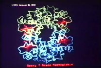
00:46:12 it was possible to construct a model
00:46:16 of the sickle cell precipitate,
00:46:20 which you see in the next picture.
00:46:23 So there it is,
00:46:25 and it consists of six filaments
00:46:31 of single hemoglobin molecules
00:46:33 wrapped around a hollow tube.
00:46:37 And the repeat from this molecule
00:46:41 to one that lies exactly over the top of it
00:46:49 is 520 angstroms,
00:46:53 and the repeat between layers is 62 angstroms.
00:46:56 So here you see the plan.
00:46:58 Another way of looking at it is
00:47:00 that you can think of it as a succession of hexagonal rings,
00:47:04 each ring being displaced relative to its predecessor
00:47:07 by 7.3 degrees.
00:47:14 So, then,
00:47:17 see, the exciting thing about this structure is this.
00:47:21 If we could resolve the nature of the contacts
00:47:24 between the subunits,
00:47:27 then we might be able to design some drug, say,
00:47:31 which competes with the site here
00:47:34 and actually prevents the aggregation.
00:47:37 So, we then ask ourselves,
00:47:43 what are the sites of contact
00:47:45 where these molecules stick together?
00:47:48 We don't know yet,
00:47:50 but the optical dichroism gives us a clue
00:47:54 at least to the orientation of the molecules,
00:47:57 and that you see in the next picture.
00:48:00 Now, you'll see a hemoglobin model
00:48:03 oriented in the way it would be in the fiber.
00:48:07 So, there are the alpha and here are the beta chains,
00:48:10 and the hemes are approximately normal to the fiber axis,
00:48:14 and so the next molecules would be there and there.
00:48:19 But now, where are the sickle cell sites?
00:48:22 One is here, and the other one is there.
00:48:26 And in this picture, it sort of looks okay.
00:48:29 You think, all right,
00:48:31 this valine might stick to some site in the next molecule,
00:48:39 but in fact, this is only because the photograph is foreshortened.
00:48:44 If you look at the actual model,
00:48:46 the valines are in here and there, you see,
00:48:50 because these things come further in,
00:48:53 and it's quite impossible to bring them into contact
00:48:57 with the next molecule above or below.
00:49:00 So, clearly, if these play a part,
00:49:04 they must be linking the molecules within the hexagon side by side,
00:49:11 and there is an observation which suggests that this is really so,
00:49:15 because having sort of found this,
00:49:18 John Finch and I wondered how unique this precipitate was.
00:49:23 So he examined oxygenated normal cells, deoxygenated normal cells,
00:49:30 oxygenated sickle cells, oxygenated and deoxygenated fetal cells,
00:49:36 and they were all empty except deoxygenated normal cells,
00:49:43 in which he found fibers looking exactly like
00:49:51 the sort of aggregates of sickle cell filaments
00:49:55 that we had seen in free solution,
00:49:58 only at a much lower concentration.
00:50:01 So the next picture shows you this.
00:50:04 Can I have...
00:50:06 Oh, can you push it up, please?
00:50:10 Something's gone wrong.
00:50:13 No, still down there.
00:50:17 Well, I'll tell you in the meantime what it is.
00:50:20 You find aggregates of these filaments in the normal cells,
00:50:25 but they are irregular.
00:50:28 You don't get the regular hexagonal structure
00:50:35 that the hemoglobin S fibers form.
00:50:39 So telling you that hemoglobin A as such also has a tendency
00:50:44 to aggregate end-to-end,
00:50:46 but apparently not having the stabilizing interaction
00:50:50 to form the 6-fold fiber, it's much less soluble.
00:50:54 But look, never mind, let's go on to the next slide,
00:50:57 if this is so recalcitrant.
00:51:00 Yes.
00:51:02 So, now, I spent a lot of time on sickle cell hemoglobin
00:51:06 because of its great importance,
00:51:08 but now I want to tell you about some of the others,
00:51:15 which admittedly occur mainly in isolated families,
00:51:19 in heterozygotes,
00:51:21 but they are most illuminating about the sort of interplay
00:51:25 between protein structure and function.
00:51:29 Perhaps I should still say this,
00:51:32 that while the sickle cell gene is very frequent
00:51:37 in some parts of West Africa,
00:51:39 it occurs in about 40 percent of the population,
00:51:43 there are only three other variants
00:51:46 which are sufficiently frequent to occur in homozygotes,
00:51:50 and none of these produce severe disease.
00:51:58 All the other 180-odd are rare,
00:52:03 occur only in isolated families,
00:52:05 so that the probability of finding heterozygotes,
00:52:09 finding homozygotes, is very small.
00:52:12 But some of them produce severe clinical symptoms
00:52:17 even in heterozygotes,
00:52:19 and I shall now show you some of the reasons why.
00:52:24 Now, the first group,
00:52:29 go back to this argument
00:52:35 about the nonpolar interior of the globin chains,
00:52:39 which has to be sealed off from water
00:52:42 in order to function properly.
00:52:44 So, there is a mutation, hemoglobin Hammersmith,
00:52:50 in which a phenylalanine,
00:52:52 which hangs down here and seals off the heme pocket,
00:52:56 is replaced by a serine that leaves it open.
00:52:59 And this causes an instability of the beta chains,
00:53:05 causes the globin to be denatured in the red cell
00:53:10 and precipitate in the form of Heinz bodies,
00:53:12 which make the cell rigid,
00:53:14 and thereby liable to early destruction.
00:53:19 So that it results in a severe anemia.
00:53:22 They actually have a girl,
00:53:24 I think about an 18-year-old girl,
00:53:27 at Hammersmith, who has this disease.
00:53:29 She has a hemoglobin of only 6%.
00:53:32 Her urine is black because the hemolytic anemia is so severe.
00:53:39 But nevertheless, apparently, she's relatively fit
00:53:43 and actually goes swimming and plays tennis,
00:53:46 which is quite remarkable.
00:53:50 Now, another mutant of the same sort of physiological effect is this.
00:53:59 Not very well visible here.
00:54:02 It's a close contact between the helices B and E.
00:54:08 This one and that one.
00:54:10 There's one point where they're so close together
00:54:13 that there's not room for a side chain between them.
00:54:18 So both the residues of the contact are glycines.
00:54:22 In hemoglobin savannah, one of these glycines,
00:54:25 in position B24, is replaced by a valine,
00:54:30 which has a bulky side chain
00:54:32 and obviously squeezes these two helices apart.
00:54:38 And again, you have the same symptom,
00:54:40 enhanced by this severe hemolytic anemia.
00:54:45 Now, another very interesting mutation is hemoglobin Wien for Vienna.
00:54:51 In here, in this crevice, lies a tyrosine,
00:54:54 130, which is part of the helix H,
00:54:57 whose phenyl group is in...
00:54:59 whose sort of benzene ring is internal,
00:55:02 but the phenolic hydroxyl makes a hydrogen bond just outside.
00:55:05 Now, in hemoglobin Wien, this is replaced by aspartate,
00:55:09 which is short and produces a polar group
00:55:12 in the nonpolar interior of the protein,
00:55:14 and this produces a severe instability
00:55:18 so that the protein precipitates.
00:55:21 So the moral then is...
00:55:23 Well, there are others.
00:55:24 There's another mutation where a leucine is replaced by an arginine,
00:55:28 which leaves the holes through which water can get in.
00:55:31 So the moral of all this is
00:55:34 if you let water into the interior,
00:55:38 you produce a deleterious mutation
00:55:41 which affects the stability and function of the protein.
00:55:46 Now, quite another class of proteins
00:55:49 producing hematological symptoms
00:55:52 are those that affect the oxygen affinity,
00:55:55 and they do this often
00:55:57 merely by affecting the relative stability
00:56:01 of the venous and the arterial form,
00:56:03 and I'll show you an interesting example.
00:56:05 Now, the next slide
00:56:08 shows us a diagram you've seen before
00:56:12 with these alternative positions
00:56:15 of the bond between the contact
00:56:18 between the alpha and the beta subunit
00:56:21 and these hydrogen bonds.
00:56:23 Now, in hemoglobin Kempsi,
00:56:25 this aspartate is replaced by an asparagine
00:56:29 so that this single hydrogen bond here
00:56:33 becomes inactive.
00:56:38 As a result,
00:56:41 this hemoglobin crystallizes in the oxy form
00:56:45 even when you have completely deoxygenated it
00:56:49 because you have shifted the equilibrium
00:56:53 of these two allosteric forms
00:56:56 by the equivalent of two hydrogen bonds
00:56:59 which may be, say, 6 kilocalories,
00:57:01 something of that order.
00:57:03 We don't know exactly what.
00:57:05 There's another one
00:57:08 should be close to your local heart's
00:57:13 hemoglobin Yakima,
00:57:15 in which this is replaced by histidine
00:57:18 and has similar physiological properties.
00:57:22 Now, so, then,
00:57:25 the patients who have this
00:57:27 have a high oxygen affinity
00:57:29 and this means
00:57:33 that the organism has difficulty
00:57:36 in drawing off oxygen
00:57:38 so through the sort of feedback mechanism
00:57:41 which you have in the kidneys,
00:57:43 the lack of oxygen
00:57:46 stimulates the synthesis of erythropoietin
00:57:51 which in turn stimulates red cell synthesis
00:57:55 resulting in polycythemia.
00:57:57 So then these patients have polycythemia
00:58:00 merely through a disturbance
00:58:03 of the allosteric equilibrium
00:58:05 between these two forms.
00:58:07 Now, the converse you see there,
00:58:09 there's a hemoglobin Kansas
00:58:11 in which this asparagine
00:58:13 is replaced by a threonine
00:58:16 which cannot make that hydrogen bond.
00:58:19 So hemoglobin Kansas
00:58:22 has this structure
00:58:24 even when it's fully oxygenated
00:58:26 and the oxygen affinity is low therefore
00:58:31 and resulting in aplastic anemia.
00:58:34 So again, the anemia is due merely
00:58:37 to too much oxygen being available,
00:58:40 oxygen being drawn off too easily
00:58:43 so that the synthesis of erythropoietin
00:58:46 is repressed
00:58:48 and the anemia results.
00:58:57 There are two other hemoglobin,
00:59:01 abnormal hemoglobins
00:59:05 discovered here.
00:59:08 Let's say one of them discovered here,
00:59:10 both of them analyzed in great detail here.
00:59:13 These are hemoglobin Rainier and Bethesda.
00:59:17 Can I have the next slide, please?
00:59:20 They both have very high oxygen affinities.
00:59:25 Here you see the oxygen equilibrium curves
00:59:28 of normal hemoglobin
00:59:30 at three different pHs.
00:59:33 So this would be at low pH
00:59:37 and that one.
At the inaugural Philip Wilcox Memorial Lecture at University of Washington, Max Perutz presents his research on the structure and function of hemoglobin, some mutations that can occur in the protein and their physiological effects.
The quality of the original tape has unfortunately degraded over time, resulting in image and audio issues. This is an incomplete recording and does not contain the end of the lecture.
| Property | Value |
|---|---|
| Creator of work | |
| Place of creation | |
| Format | |
| Genre | |
| Medium | |
| Extent |
|
| Language | |
| Subject | |
| Rights | In Copyright - Rights-holder(s) Unlocatable or Unidentifiable |
| Credit line |
|
Institutional location
| Department | |
|---|---|
| Collection | |
| Series arrangement |
|
| Physical container |
|
Related Items
Cite as
University of Washington, and Max F. Perutz. “Molecular Pathology of Human Hemoglobin.” Dvd, u-matic (tm), June 1973. History of Molecular Biology Collection, Box 95, Folder 12. Science History Institute. Philadelphia. https://digital.sciencehistory.org/works/7jt02wb.
This citation is automatically generated and may contain errors.