Species Adaptation in the Hemoglobin Molecule
A Seminar by Max Perutz
- 1985-Mar-20
These captions and transcript were generated by a computer and may contain errors. If there are significant errors that should be corrected, please let us know by emailing digital@sciencehistory.org.
Transcript
00:00:50 It's my pleasure, a big pleasure to welcome to the University of Chicago today Dr. Max Perutz of the MRC Laboratory of Electrobiology in Cambridge.
00:01:13 Some people are said to be of such distinction that they need no introduction, if ever that were the case, it's true here.
00:01:21 However, it's a really good opportunity to introduce anyone of Max's distinction, I'm going to introduce him anyway.
00:01:30 Max started his scientific career as an undergraduate in chemistry at the University of Vienna, and in 1936 went to Cambridge to take up graduate school.
00:01:41 Now he originally wanted to work on amino acids with Colin Hopkins, but his mentor in Vienna, Herman Mark, pre-polymer chemist,
00:01:50 had just returned from Cambridge and was all aglow with reports of X-ray diffraction patterns from crystals of protein,
00:01:59 having been taken by J.D. Bernal and Dorothy Crowfoot, subsequently Dorothy Crowfoot Hodgkin, and their collaborators in Cambridge.
00:02:09 They were so excited about the prospect of being able to visualize protein molecules in detail so that they could figure out how they worked,
00:02:19 and they transmitted this enthusiasm to Mark, who signed Max up to work with Bernal.
00:02:26 Not that Max knew about this until he paid a call on him.
00:02:31 So Max went to Cambridge and began his work there on protein crystallography, and he began working on the hemoglobin molecule.
00:02:42 Well there was a lot of intervening history and a lot of hard work, nineteen- in the war years as well.
00:02:48 We'll jump ahead to 1947, when Max became director of the MRC unit at the Cavendish lab.
00:02:56 He had a giant staff of one person, John Kendrew.
00:03:00 Well the lab grew, and you all know the luminaries who came there.
00:03:05 And in 1962 they moved to their new residence on Hills Road with a much expanded staff.
00:03:14 In 1962 their work was acknowledged by the laboratories winning four Nobel Prizes in one year.
00:03:21 Max and John Kendrew in chemistry for their work on the hemoproteins, the crystallography of the hemoproteins,
00:03:27 and some young fellows by the name of Watson and Crick in collaboration with Wilkins for the structure of DNA.
00:03:35 Well since that time that laboratory has manifested itself in a manner that I believe no others have in the area of biology, certainly at the molecular level.
00:03:48 Having won in total seven Nobel Prizes to a building about half the size of this one, isn't that quite an accomplishment?
00:03:56 And it's been under the directorship of Max.
00:03:58 Meanwhile, about five years ago Max turned over the directorship to Sydney and it was a kind of revert.
00:04:05 He re-entered into his already active program on hemoglobin structure-function relationship.
00:04:10 He's going to tell us today about one of the areas that he's been studying and that's the species adaptation in the hemoglobin molecule.
00:04:40 Can you hear me at the back?
00:05:09 Yes.
00:05:10 Yes, good.
00:05:13 Levantin complained in an article in the Scientific American a few years ago that it has proved remarkably difficult
00:05:23 to get compelling evidence for changes in enzymes brought about by selection, not to speak of adaptive changes.
00:05:32 Fortunately, such evidence has recently been gathered for hemoglobin, whose response to different chemical stimuli varies widely in vertebrates living in different environments.
00:05:46 So one asks oneself, how did this adaptation come about?
00:05:50 Is it the result of changes in tertiary or paternal structure of the hemoglobin molecule?
00:05:57 Or has it been brought about by the gradual accumulation of minor mutations, each producing a small change in chemical affinity?
00:06:05 Or there are fewer amino acid substitutions in T-positions, each producing a large change in chemical affinity.
00:06:13 Have similar changes in affinity in different animal species been brought about by the same amino acid substitutions or the different ones?
00:06:25 And finally, are most amino acid substitutions between species functionally significant?
00:06:35 And have they evolved by Darwinian selection?
00:06:41 Or are they caused by random fixation of neutral or almost neutral mutations?
00:06:46 Are they results merely of genetic drift?
00:06:51 Now, by studying the adaptation of the hemoglobin molecule, one can at least begin to answer some of these fundamental questions.
00:07:01 Now, perhaps I should introduce hemoglobin, because we won't all be familiar with it.
00:07:09 And for this purpose, I would like to have the first stereoslide.
00:07:18 Have you all got stereoglasses, or stereoglasses have your neighbors got them?
00:07:31 What about these? Are these pairs?
00:07:35 Yes, I think you do have a couple of them.
00:07:40 So, this shows a...
00:07:50 Not very clearly visible from the side.
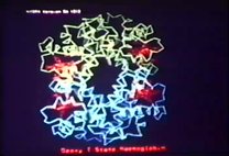
00:07:54 This shows the stereo view of my own hemoglobin model, which in its four subunits,
00:08:04 the two white subchains representing the alpha chains and the black ones, the beta chains,
00:08:12 and the hemes being in the pockets on the surface of the molecule.
00:08:19 And here is the position of the reactive start-hybrid group of hemoglobin,
00:08:25 at residue 93 beta, which we shall be talking about.
00:08:30 And going through the center of the molecule from its top, is a two-fold symmetry axis,
00:08:36 so that a rotation of 180 degrees brings this black chain of the congruence to its opposite.
00:08:47 The most important feature of this molecule is its change of structure on taking up oxygen.
00:08:59 This is the oxy-hemoglobin structure.
00:09:02 On loss of oxygen, one pair of subunits, one white and one black chain,
00:09:09 rotates relative to the other pair by 15 degrees,
00:09:13 thus widening the slit between the two beta chains at the bottom
00:09:18 and opening a binding site for the allosteric regulator 2,3-biphosphoglycerate.
00:09:24 So, hemoglobin alternates between two structures,
00:09:28 one with an oxy- or T-structure with a low oxygen affinity
00:09:32 and a high affinity for protons, chlorides, carbon dioxide,
00:09:37 and in mammals 2,3-biphosphoglycerate,
00:09:41 the other structure in which these relative affinities are reversed.
00:09:48 The oxygen affinities of the two alternative structures in mammals
00:09:55 differ by the equivalent of 3 kilocalories per mole,
00:10:01 and that is usually referred to as the free energy of heme-heme interaction.
00:10:06 So, so much for this introduction,
00:10:09 and now we can stop the stereos and look at the first monoslide, please,
00:10:17 which shows the tertiary structure of the hemoglobin molecule
00:10:25 and the notation that is used to designate the different residues.
00:10:32 This was introduced by Kangu from myoglobin and has been very useful
00:10:37 because the number of amino acid residues in the hemoglobin of different animal classes differs,
00:10:45 say, the alpha chain in human hemoglobin has 142 residues and in fish 140 - sorry, in humans 141 and in fish 142,
00:10:57 but insertions and deletions occur at the corners of non-helical segments
00:11:05 and the number in the helical segment tends to be constant.
00:11:09 So we call the helical segments A, B, C, D, etc. to H,
00:11:15 and the non-helical one, the first one we call NA for the segment with the amino end,
00:11:25 and then C, D, and so on until we come to GH here,
00:11:31 and finally to HC, which designates several non-helical residues at the carboxyl end.
00:11:41 The heme you see in this pocket here is sandwiched between two histidines,
00:11:47 one covalently linked to the R, which is residue F8,
00:11:52 to its residue along helix F,
00:11:55 and the other, the distal one, which is hydrogen bonded to the linked oxygen when it's there,
00:12:04 which is E7, so it's the seventh residue along the E helix.
00:12:10 The next slide shows you a view of the hemoglobin molecule,
00:12:16 the same one that we saw before,
00:12:19 from the, along the direction of the super-extremity axis,
00:12:24 which runs through the central cavity as you see here,
00:12:28 and there you see the amino termini of the alpha chain,
00:12:33 which have some important part to play in function.
00:12:40 Right, now, the next slide shows you a logarithmic plot
00:12:47 of two pythagorean-oxygen equilibrium curves of human hemoglobin
00:12:51 across on the x axis, the partial pressure,
00:12:54 the log of the partial pressure of oxygen on the ordinate,
00:12:58 the log of Y over 1 minus Y,
00:13:02 where Y is the fractional saturation with oxygen.
00:13:07 So these equilibrium curves are sigmoid,
00:13:11 because the binding of oxygen is cooperative,
00:13:15 and they are pH-dependent,
00:13:17 so that the oxygen affinity rises with increasing pH,
00:13:26 with lowering hydrogen ion concentration,
00:13:31 so oxygen and hydrogen ions are antagonistic,
00:13:35 and there's a profound physiological purpose for this,
00:13:41 because hemoglobin is a two-way respiratory carrier,
00:13:45 it carries oxygen from the lungs to the tissues,
00:13:48 and it promotes the return transport of carbon dioxide
00:13:52 from the tissues back to the lungs,
00:13:54 and it does that by picking up hydrogen ions on loss of oxygen.
00:14:02 From loss of oxygen, hemoglobin mops up the hydrogen ions
00:14:07 that are liberated in the reaction of carbon dioxide and water
00:14:12 to form bicarbonate,
00:14:14 thus shifting the mass equilibrium towards bicarbonate ions,
00:14:20 which are soluble and can be transported back to the lungs in the serum.
00:14:26 In the lungs, hemoglobin takes up oxygen,
00:14:32 liberates hydrogen ions,
00:14:35 which pushes the equation back to the left
00:14:39 towards carbon dioxide and water,
00:14:42 and carbon dioxide is exhaled.
00:14:44 But moreover, you see that the liberation of lactic and carbonic acids in the tissues
00:14:52 lowers the oxygen affinity
00:14:54 and so promotes the liberation of oxygen.
00:14:58 Now, this effect of the H-dependent of the oxygen equilibrium
00:15:06 and the antagonistic effects of oxygen and hydrogen ions
00:15:12 are known as the Bohr effect,
00:15:14 after Christian Bohr,
00:15:16 and many physiologists who discovered them.
00:15:21 Now, so much then for the human.
00:15:36 Some years ago, an Italian chemist, Maurizio Brunori,
00:15:45 showed me what the equivalent curves are in the trout.
00:15:50 Can I have the next slide, please?
00:15:54 So, here again, you see we have the log of the partial pressure of oxygen,
00:15:59 and instead of y, it holds the fractional separation x bar,
00:16:04 but it's the same thing.
00:16:06 And here are the equilibrium curves.
00:16:12 Note that only at this middle pH, which is 7.15,
00:16:19 does the equilibrium curve resemble the human one.
00:16:26 At alkaline pH, it's no longer sigmoid,
00:16:31 and at acid pH, it becomes side-basic,
00:16:35 and the shift is far to the right,
00:16:38 so that the influence of pH on the equilibrium curve
00:16:49 is enormously exaggerated in this trout hemoglobin.
00:16:56 But that's not all.
00:16:58 Trout actually has two kinds of hemoglobin,
00:17:01 one which is enormously dependent,
00:17:04 and another with a sigmoid oxygen equilibrium curve
00:17:08 that is totally independent of pH and all the other co-factors
00:17:12 which shift the curve in human hemoglobin.
00:17:18 Well, Brunori, with an old friend, came along and said,
00:17:23 how do you explain this?
00:17:25 And I said, I can't do it if you don't show me a sequence.
00:17:31 Now, before I show you the sequence,
00:17:36 let me explain what its use is to the fish.
00:17:44 The effect to which Brunori drew my attention
00:17:48 was originally discovered by an American geologist,
00:17:52 Ruth, working at the Duke University
00:17:56 marine biology laboratory in Beaufort, North Carolina,
00:18:01 in the 1930s,
00:18:04 and who showed that fish use this
00:18:09 to fill their swim bladder with oxygen.
00:18:13 My next slide shows you a diagrammatic sketch
00:18:18 of the anatomy of the swim bladder
00:18:22 which has attached to it what is called here a secretory epithelium,
00:18:29 in effect, a gland which secretes lactic acid into the blood
00:18:34 and so acidifies it.
00:18:37 And because the oxygen affinity is so strongly pH-dependent,
00:18:45 the secretion of lactic acid leads to the liberation of oxygen in the swim bladder.
00:18:51 But again, there's more to it, because attached to the swim bladder
00:18:56 is a beautiful counter-current system of small capillaries
00:19:00 where a gradient of increasing lactate concentration
00:19:09 is converted to a gradient of decreasing oxygen saturation of the hemoglobin.
00:19:16 In other words, this is a device for transferring the unused oxygen of the veins to the arteries
00:19:30 so that more of the oxygen that is carried can be secreted into the swim bladder.
00:19:38 The next slide shows you a microscope section through this organ
00:19:46 and you see that, well, you can't see it because it is closed,
00:19:52 that each of these arteries tends to be surrounded by veins
00:19:57 and vice versa so that you get a perfect exchange.
00:20:03 Now, since then people have discovered that there is another such system
00:20:08 at the back of the retina of fish ensuring secretion of oxygen into the eye.
00:20:17 So the fish use this rudiment then both for regulating their buoyancy
00:20:28 and improving their vision.
00:20:33 So, as I told you, Brunori asked me how do you explain this
00:20:40 and at the fullness of time the amino acid sequences of several fish hemoglobins
00:20:45 became known and were [carved as the goldfish] of these two components of trout hemoglobin
00:20:54 and of an American fish called [fox].
00:20:59 So, I looked at these and they seem very unpromising
00:21:10 because about half the amino acid residues of human hemoglobin differ from the fish.
00:21:20 The hemoglobin amino acid sequences are surprisingly variable.
00:21:27 You know, if you look at all the hemoglobins of vertebrates and de-vertebrates
00:21:33 there are in fact only two invariant residues.
00:21:36 One is the proximal histidine which is essential for the oxygen binding
00:21:42 and the other is the phenylalanine which is in the fish's [pockets].
00:21:46 And all the other residues are exchangeable.
00:21:50 So, in the differences between trout and human, there is, as I say, about 140.
00:21:59 So where do you start?
00:22:01 Well, as often in science, you have to take your courage into your hand
00:22:08 and decide you're going to look at these amino acid mutations one by one
00:22:13 and ask yourself for each one of them whether that could possibly have produced
00:22:19 this root effect, this enormous increase in the pH dependence of the oxygenic hemoglobin.
00:22:26 And when you do that, you find that the vast majority of the mutations are in fact conservative ones
00:22:32 of non-polar residues in the interior or polar residues in the exterior
00:22:39 which clearly have no particular function.
00:22:42 But then one particular replacement caught my eye, which I now want to tell you about.
00:22:48 And this is of the reactive cysteine in mammals by serine in all these fish.
00:22:58 The next slide shows you just the position of the reactive H-group again,
00:23:07 which is here in the model.
00:23:09 And in the next slide, the function of the human hemoglobin.
00:23:18 So, here you have a diagrammatic sketch showing the heme and the human deoxyhemoglobin
00:23:24 and the human oxy.
00:23:26 The deoxy, the R is out of the frame of the forefront.
00:23:30 Here's the proximal histidine residue, F8.
00:23:34 And the next one, F9, is this reactive cysteine,
00:23:39 which in deoxyhemoglobin is surrounded by the C-terminal histidine
00:23:45 that forms a salt bridge with aspartate
00:23:52 and with the lysine C-prime alpha.
00:24:00 Now, in human oxyhemoglobin, the salt bridge is broken
00:24:06 and the C-terminal histidine instead forms a salt bridge with the lysine here.
00:24:15 The idazole accepts the hydrogen bond from its own end, which is not yet shown here.
00:24:24 So, this changing conformation of the C-terminal histidine
00:24:31 turns out to be responsible for about half the Bohr effect in human hemoglobin
00:24:37 because salt bridge with aspartate reduces decay to eight
00:24:43 and the rupture of the salt bridge lowers its decay to about seven.
00:24:53 Right. Now, what I noticed then was that in restriction hemoglobin,
00:24:58 this SH group is replaced by serine.
00:25:03 In human hemoglobin, it seems to act merely as a spacer.
00:25:13 That is to say, the cysteine SH forms on the very weak hydrogen bond.
00:25:22 For instance, if you look at the neutron refraction structure of cysteine itself,
00:25:28 then the SH group is in contact with the carboxylate of a neighboring cysteine molecule,
00:25:37 but the distance from the hydrogen to the oxygen is no larger than the sum of the Van der Waals radii.
00:25:44 Whereas if you did the same with a serine,
00:25:47 you'd probably find very strong hydrogen bonding,
00:25:50 the distance being several tens of angstroms shorter than the sum of the Van der Waals radii.
00:25:56 Now, the next slide shows you the position in carp,
00:26:02 where, as I said, we have a serine in this position.
00:26:07 So, what I did was take the model of human hemoglobin
00:26:12 and simply replace the cysteine by a serine.
00:26:17 And when I did that, I found that the serine can donate the hydrogen bond to this terminal carboxyl,
00:26:26 one that would be oxygen that would not bond with the live thing,
00:26:30 and that it can accept the hydrogen bond from the NH of this T-terminal histidine
00:26:38 in the deoxy or T-structure.
00:26:41 In the oxy or R-structure, these bonds would be broken.
00:26:45 So, I argued then that if [when the] salt bridge is closed,
00:26:50 these bonds would contribute something between 1 and 2 kilocalories per mole of stabilization energy to the T-structure,
00:27:04 and this may be responsible for the enormously reduced oxygen affinity and acid pH,
00:27:15 because acid pH stabilizes that sulfate.
00:27:19 Well, that was just an idea, but how do you find out whether there is anything in it?
00:27:28 Well, one way in which Kilmartin had tested,
00:27:42 and my colleague Kilmartin had tested the original Bohr mechanism,
00:27:48 was to leave away the T-terminal histidine,
00:27:54 and see how it affected the Bohr effect, and the T-terminal capacity.
00:28:00 Now, in carp, this was much more difficult,
00:28:03 in fish hemoglobin because you can't stick anything into alpha and beta chains,
00:28:07 but Lawrence Parker at the University of Lincoln in Nebraska succeeded in doing this,
00:28:13 he cleaved away the T-terminal histidine,
00:28:16 and measured the influence of that cleavage on the oxygen equilibria.
00:28:26 Now, the next slide, please.
00:28:29 So, here the full curve of naked carp hemoglobin,
00:28:38 and there you see the curve at pH 7, and at pH 6, and pH 9,
00:28:50 and the working curves show that this is carp hemoglobin,
00:28:56 in which the root effect, Bohr effect is greatly reduced.
00:29:04 So, this is a kind of negative test, but I hope that we can soon have a positive one.
00:29:14 It comes from my colleague Kiyoshi Nagai,
00:29:22 has developed a method of cloning human hemoglobin in E. coli,
00:29:28 so that we can now subject it to a redirected mutagenesis,
00:29:33 and convert the cysteine in position 8-9 to a serine,
00:29:41 and see what influence it will have on the oxygen equilibria.
00:29:46 So, in the fullness of time, I hope that we will see a communication,
00:29:53 in Nature, on turning man into fish,
00:29:57 and see what the result of this experiment is going to be.
00:30:04 So, this then is one of the adaptive mechanisms.
00:30:12 Now, another one, for reasons which are not so clear, is this.
00:30:22 In human hemoglobin, the oxygen affinity is regulated by 2,3-diphosphoglycerate.
00:30:29 So, the next slide shows you the binding site in human hemoglobin.
00:30:36 There is the diphosphoglycerate, which is phosphate, which is carboxylate,
00:30:41 and in deoxyhemoglobin, there is a constellation of 8 complementary cationic groups,
00:30:49 which bind to the diphosphoglycerate anion.
00:30:53 So, you see these two histamines, these two lysines, this alpha-amino group.
00:31:00 In the transition to the oxygen structure, these two segments, the ES segments,
00:31:09 move apart, move together, and the alpha-amino groups move apart,
00:31:15 so that the complementarity is lost, and the DPP dissociates.
00:31:19 The next slide shows you the situation in oxyhemoglobin.
00:31:25 You see these two valines are now much further apart,
00:31:30 and the lysines are closer together.
00:31:33 And here you see another way to do lysine, a hundred and forty-four.
00:31:39 So, human hemoglobin responds strongly to diphosphoglycerate,
00:31:47 but very weakly to ATP or GTP.
00:31:52 In fish, it is reversed.
00:31:54 They show a very strong response to GTP, about half that response to ATP,
00:32:01 and a very weak one to diphosphoglycerate.
00:32:05 So, in the red cells, there is a very high concentration of the two triphosphates,
00:32:13 purine triphosphates, ATP and GTP.
00:32:17 And so, again, the physiologist asks me,
00:32:22 how can you convert these affinities?
00:32:26 What can reverse the relative affinities,
00:32:31 or change it from diphosphoglycerate to purine triphosphate?
00:32:37 So, again, the sequence gives a clue.
00:32:43 The sequence shows that in these fish hemoglobin,
00:32:47 this histidine 2 here is referred to.
00:32:51 It's replaced either by an aspartate or a glutamate.
00:32:55 We have an acid here.
00:32:57 And this histidine 143 is replaced by a stronger base,
00:33:03 by arginine.
00:33:06 Moreover, Allen Edmondson showed many years ago
00:33:10 that treatment of the alpha-amino group entirely abolishes the affinity for ATP.
00:33:22 So, the alpha-amino groups are clearly involved.
00:33:26 Right.
00:33:27 Now, again, I said, and this is a crucial point,
00:33:31 I said, let us assume that the tertiary-quaternary structure
00:33:36 of 4-carbon hemoglobin is exactly the same as that of human hemoglobin.
00:33:42 And merely replace one side chain by another.
00:33:50 We merely remove this histidine by a glutamate,
00:33:54 that histidine by arginine,
00:33:57 and then introduce a molecular model of ATP and see whether it fits.
00:34:04 Now, may I have the next stereo slide, please?
00:34:26 So, what have we got here?
00:34:29 Here's the alpha-amino group of the valine 1-alpha.
00:34:35 Here's this glutamate 2.
00:34:38 And there's lysine 82, which is conserved.
00:34:42 And here's the arginine, which was 143 in humans.
00:34:50 So, what I did is I simply took an extended model of ATP
00:34:56 and put it purely in the anti-conformation.
00:35:00 I said, we must have the gamma phosphate linked to the valine 1-alpha-amino group
00:35:11 as our experiment would have worked if it did.
00:35:16 And clearly, the glutamate [also had to be bound]
00:35:23 with the amino group of the adenine.
00:35:26 And so that's all I did, and it fitted in perfectly.
00:35:33 Now, next, I wondered how could I fit in TTP,
00:35:44 and how could I explain that the affinity of TTP,
00:35:49 the binding constant, is twice that of ATP.
00:35:58 And first, I was perplexed, because in TTP,
00:36:04 this amino group here is replaced by a carbonyl.
00:36:08 But then, with the help of an NMR experiment,
00:36:12 which I shall show you presently, I dropped the [fluid]
00:36:18 and simply turned this model.
00:36:22 Well, I fixed a guanine here and turned the entire model
00:36:27 by 180 degrees about this long axis.
00:36:32 And the next slide shows you the result.
00:36:37 May I have the next stereo
00:36:39 please?
00:36:45 So this needs adjusting, so wait a little bit before you look at it.
00:36:53 I think, isn't it a long?
00:36:56 It's vertical adjustment.
00:36:57 It's vertical adjustment.
00:36:58 It's a long way out of space.
00:37:01 Adjust it.
00:37:05 That's better.
00:37:06 Now it's right.
00:37:07 Good.
00:37:08 So, here you see the TTP again, and you see that
00:37:22 its amino group forms a hydrogen bond again with glutamate.
00:37:29 And this time, one of the hydroxides of the ribose
00:37:35 is in a position to form a hydrogen bond
00:37:39 with the alpha-amino group of the valine.
00:37:42 So the TTP is bound by one additional hydrogen bond,
00:37:47 and this accounts, lastly, for its greater affinity
00:37:52 compared to TTP.
00:37:55 Right, thank you.
00:38:04 So, can we have the next mono slide?
00:38:09 In fact, the job wasn't as easy as that.
00:38:13 And my first proposal for the TTP binding
00:38:16 was shown to be incorrect by NMR, [as started].
00:38:24 I collaborated with two young NMR specialists
00:38:28 at the National Institute of Medicine in London,
00:38:31 Marius Clore and Angela Gronenborn,
00:38:34 and they determined the combination of the bound ATP and TTP
00:38:43 by the time-dependent transferred nuclear overhauling effect,
00:38:49 which depends on a fast exchange between free and bound cofactor.
00:39:00 You get, what you do is you irradiate the resonance of one
00:39:07 of the hydrogens and determine the interaction
00:39:14 with another hydrogen as a function of time.
00:39:21 So down here, you see the notation for ATP,
00:39:28 and what we wanted to know was this.
00:39:31 We wanted to know whether the corrin ring is
00:39:34 in anti- or syn-position relative to the rival.
00:39:39 The way you see it here, it is in anti-position,
00:39:42 but if you turn it by 180 degrees,
00:39:45 about this ends in bond, which would be syn.
00:39:48 Sorry, is it in syn?
00:39:52 Yes, I think you were right, it's in syn.
00:39:57 So, what we did then was to irradiate the H1 prime,
00:40:06 that would be this proton,
00:40:09 and then determine its effect on the resonance of H8,
00:40:16 which you see here, and then irradiate the H2 prime,
00:40:23 determining the resonance of each dehydrogen here,
00:40:29 dehydrogen there, and the H1 prime, and so on.
00:40:36 So the object was to see whether it was the hydrogen at C2
00:40:44 or the hydrogen at C8, which was next to the hydrogen
00:40:49 of the ribose ring.
00:40:52 Now, when the hydrogens are close together,
00:41:00 the curve is convex like that.
00:41:05 When they are far apart, it is concave like this.
00:41:09 So what this experiment tells us is that H2
00:41:14 is within about 3 or 3.5 angstroms of H2 bar.
00:41:21 So like so.
00:41:23 And that it is distant from H8.
00:41:28 H3 bar is close to H2.
00:41:35 And H1, again, is close to H2, but distant from H8.
00:41:42 The result told us that the purine ring
00:41:45 is an anti-conformation relative to the rivals
00:41:50 in both ATP and GTP.
00:41:52 And this helped me to get the model right.
00:41:56 The next slide shows you another NMR experiment with P31,
00:42:01 where Clore and Gronenborn tried to find out
00:42:05 whether the phosphate chain is extended or coiled.
00:42:09 And the result showed that the resonances were similar
00:42:14 to that of 3-ATP, in which the chain is known to be extended
00:42:19 so that I got the conformation for that.
00:42:26 Now, I went into this, you see, to stress the point
00:42:30 that it is possible for a protein to change its affinity
00:42:38 from one protactor to a totally different protactor
00:42:43 without any alteration in chemical structure,
00:42:46 but merely by the replacement of a very few amino acids
00:42:50 in key positions, such as histidine 2,
00:42:54 which bound the internal phosphate of GTP
00:42:58 by glutamic acid, which instead formed a hydrogen bond
00:43:03 with the amino groups of adenine O1.
00:43:07 All right.
00:43:09 Now, there was one [hit] on all this.
00:43:14 It fell out rather nicely.
00:43:16 And that came from the other [chunk in the urine],
00:43:21 which had shown no dependence of its oxygen affinity
00:43:25 on either hydrogen ions or organic phosphate or CO2.
00:43:29 It was totally constant.
00:43:32 And in due course, Bossa, a colleague of Brunori's,
00:43:37 and [of Ron's], his colleagues, collaborators,
00:43:41 worked out the amino acid sequences
00:43:43 of these two hemoglobins, which are called
00:43:46 TROUT1 and TROUT4.
00:43:48 Now, one point that you would wonder about,
00:43:51 why should the fish want another hemoglobin
00:43:55 that shows no pH dependence in its oxygen equilibrium?
00:43:59 Well, I mean, you can't ask them,
00:44:02 but what the comparative physiologists believe is this,
00:44:09 that the trout is a very fast-moving fish,
00:44:13 and that during fast movement,
00:44:15 the pH in its skin will drop so low
00:44:19 that it can no longer take up enough oxygen
00:44:24 with this hemoglobin 4,
00:44:27 whose oxygen affinity drops so low with acid.
00:44:32 So the fish needs another hemoglobin in reserve,
00:44:36 which allows him to pick up oxygen under these conditions.
00:44:42 Now, then, you see, the question is,
00:44:47 if my ideas are right,
00:44:52 then all these key residues should be replaced
00:44:56 by, as it were, neutral ones in the TROUT1.
00:45:00 And I think the next slide shows
00:45:05 the amino acid sequences of human hemoglobin
00:45:11 in these key positions compared to trout.
00:45:16 So, first of all, let's look and see
00:45:20 what the C-terminal histidine does,
00:45:23 which contributes powerfully to the Bohr effect.
00:45:31 So it's histidine in human, and histidine in TROUT1,
00:45:34 but it's phenylalanine in TROUT4.
00:45:38 So there, you have only a van der Waals interaction
00:45:44 between the phenyl side chain and the helix H
00:45:52 to stabilize the key structure, but no hydrogen bond.
00:45:57 Now, then, there is this cysteine, F9,
00:46:07 which is here in TROUT4,
00:46:10 which is changed to alanine in TROUT1.
00:46:14 Then there's aspartate,
00:46:17 that forms the hydrogen bond with the C-terminal histidine,
00:46:20 that's the aspartate ST1,
00:46:22 that's glutamate in TROUT4,
00:46:25 but asparagine, an un-TROUT residue, in TROUT1.
00:46:31 So, so much, then, for the residues responsible for the root effect.
00:46:35 Now, let's look at those responsible for the binding of ATP.
00:46:40 A central role is played by this lysine, PH6.
00:46:45 You know, that's the one that's conserved in human and fish.
00:46:49 And we see in TROUT1 that's replaced by leucine.
00:46:53 The arginine, which was H21,
00:46:59 that's the one that was histidine in human,
00:47:02 arginine in TROUT4,
00:47:04 has been replaced by serine.
00:47:07 So, these two replacements are sufficient to account
00:47:13 for the loss of ATP activity in the TROUT1.
00:47:19 And that's all encouraging.
00:47:22 Now, there are other fish which have no swim bladder.
00:47:28 You see, the shark has no swim bladder,
00:47:31 it has to swim always to keep afloat.
00:47:38 If the shark stops swimming,
00:47:41 it drops to the bottom and can never swim.
00:47:46 And so, now, the shark hemoglobin
00:47:50 has, as you would expect from that,
00:47:53 only a weak Bohr effect.
00:47:55 There's no root effect right now.
00:47:57 So you ask yourself, how does this come about?
00:48:00 Well, first of all, the absence of the root effect
00:48:03 is explained by residue F9 being alanine.
00:48:08 You see?
00:48:10 So, no arginine bonds there.
00:48:14 And then, there is the C-terminal residue in histidine,
00:48:23 but there's now another residue, a lysine,
00:48:25 one term down, one thermophilic star from HB1,
00:48:30 to compete with the histidine for the hydrogen bond
00:48:35 which gets intubated,
00:48:37 so that it only weakens the Bohr effect.
00:48:42 And then, there's the lionfish, you know?
00:48:45 So the lionfish snaps up air, you see,
00:48:50 and absorbs it through its lungs,
00:48:54 so it uses its lungs for buoyancy,
00:48:58 so it doesn't have to need a swim bladder.
00:49:00 So again, we have no root effect.
00:49:05 Now, but if this...
00:49:09 Let's see how does this work.
00:49:12 If you have some therine here,
00:49:15 if you again have a second histidine there,
00:49:19 so this is H6,
00:49:21 to compete with the histidine there
00:49:24 for the solvent which can neutralize it.
00:49:32 So much then for the fish.
00:49:37 Now, what about amphibia?
00:49:42 You see, they don't really need a strong Bohr effect
00:49:48 because the pressure of carbon dioxide,
00:49:55 the blood of amphibia is so much lower
00:49:59 than in mammals, in terrestrial animals.
00:50:03 In humans, the PCO2 in the blood is 40,
00:50:08 in the human's blood is 40 millimeters, 44.
00:50:12 40 millimeters is quite big.
00:50:14 But in this amphibia,
00:50:17 it's only between 2 and 4 total
00:50:21 because of carbon dioxide,
00:50:24 that diffuses through their skin.
00:50:29 And so they, even if they live in water,
00:50:36 they don't, sorry,
00:50:38 so usually they don't have a very strong Bohr effect.
00:50:43 Xenopus, there is an exception.
00:50:46 And in the tadpoles,
00:50:49 the Bohr effect is actually reversed
00:50:52 so that the oxygen actually decreases with,
00:50:59 sorry, increases with increasing pH,
00:51:03 we see again, which we come with
00:51:05 in the case of phenylalanine.
00:51:09 Right, now, some years ago,
00:51:14 another one of these comparative physiologists
00:51:18 came along, this time a German, Christian Bauer,
00:51:21 who was in the habit of walking into an aquarium
00:51:26 and taking crocodiles by the tail,
00:51:29 pulling them into a plastic bag,
00:51:31 anaesthetizing them,
00:51:33 taking a blood sample from them
00:51:36 and measuring their oxygen equilibrium levels.
00:51:39 And by doing that,
00:51:40 he discovered that crocodilian hemoglobins
00:51:44 do not respond to any of the flow factors
00:51:47 that other vertebrates respond to,
00:51:49 but only to bicarbonate ions,
00:51:52 which has a dramatic effect on a crocodile's equilibrium.
00:51:56 So if I can have the next slide, please.
00:52:00 This shows you a comparison of the response
00:52:07 to carbon dioxide of horse hemoglobin,
00:52:10 the two broken lines,
00:52:12 and of canine hemoglobin.
00:52:14 So, in the crocodilians,
00:52:18 that really the simplest reciprocating action
00:52:21 between oxygen and the end product of oxidative metabolism
00:52:25 that you, nature, could have designed.
00:52:28 And you wonder why nature hasn't actually used
00:52:32 that very simple method anywhere else.
00:52:34 You see, so here, oxygen expels bicarbonate,
00:52:41 and bicarbonate ion expels oxygen directly.
00:52:46 And you may wonder why the crocodilians want this.
00:52:52 Now, the explanation apparently is
00:52:55 that when a crocodile decides to eat you,
00:52:59 it does not tear you to pieces,
00:53:02 but he holds you underwater until you are drowned,
00:53:06 and then eats you up at his leisure.
00:53:09 Because he can stay underwater much longer than you can.
00:53:14 A crocodile can stay submerged for as much as an hour.
00:53:18 And so, he can do this
00:53:23 because this physiology of his hemoglobin
00:53:29 enables the crocodile to just extract
00:53:33 the last bit of oxygen from its blood.
00:53:40 Whereas a human usually can't extract
00:53:44 more than about a third of the oxygen.
00:53:48 There are other mechanisms.
00:53:50 Crocodilians can also cut off the circulation from their limbs
00:53:56 and only their brain and their viscera are oxygenated.
00:54:02 But that's another matter.
00:54:04 So, the question was, how does this work?
00:54:07 Now, can we go back a few slides, please?
00:54:12 Yes, further. Further. Further. Further.
00:54:18 Right.
00:54:21 The amino acid sequence is determined
00:54:24 by Leclerc and Schneck and Boissel
00:54:28 together with Raditz and Junig
00:54:31 showed the following replacement.
00:54:34 This histidine here was replaced by a proline
00:54:41 and the valine one by a serine.
00:54:45 The lysine was preserved.
00:54:48 The histidine replaced by a serine
00:54:52 and finally the glycine 144,
00:54:56 which is a variant of mammals,
00:54:58 was replaced by glutamate.
00:55:04 Christian Bauer, the German physiologist who did that work,
00:55:08 found that the crocodilian deoxy was a response
00:55:13 to bicarbonate ions per tetramer.
00:55:16 So, he set about the job in exactly the same way as with ATP.
00:55:21 He took the human hemoglobin model,
00:55:24 checked it rigidly,
00:55:26 nearly replaced the key residues to the proline here,
00:55:31 which made the chain turn by right angle
00:55:34 and put the N-terminal residue out there,
00:55:39 replaced this lysine here by glutamate
00:55:45 and this histidine by serine.
00:55:49 So, the last stereo slide shows you the result.
00:56:16 Here is a bicarbonate ion
00:56:20 and over here is the other.
00:56:25 So, you see, I take this replacement,
00:56:28 I put in two bicarbonate ions
00:56:30 and what I found, to my surprise,
00:56:33 was that one bicarbonate oxidant
00:56:39 could accept hydrogen bonds from the serine OH
00:56:43 in the alpha-amino group.
00:56:45 The second oxidant could accept the hydrogen bonds from the lysine
00:56:51 and the third could connect the hydrogen bonds
00:56:54 to the glutamate that had replaced lysine-144.
00:57:01 So, you see, again, by doing nothing to the model
00:57:05 but replacing these two key residues,
00:57:09 I was able to turn a diphosphoglycerate binding site
00:57:17 into a pair of binding sites for two bicarbonate ions.
00:57:40 Thank you.
00:57:43 Now, the patterns in mammals.
00:57:46 The most striking adaptive phenomena you can see
00:57:50 are adaptation to high altitude.
00:57:53 Can we again go...
00:57:55 Yes, at this slide, fine.
00:57:57 In the camel and the llama,
00:58:00 two closely related species,
00:58:03 but the llama, living at high altitude,
00:58:07 has a hemoglobin with high oxygen activity
00:58:10 and you ask yourself why.
00:58:13 Can we go back one more slide, please?
00:58:17 It turns out that the camel has a glutamine here
00:58:23 which can reach it nicely
00:58:26 to form a hydrogen bond with the phosphate,
00:58:29 but the llama has an asparagine
00:58:31 whose side chain is shorter than 1.3 angstroms
00:58:35 so that it just does not quite reach.
00:58:39 Hence, the affinity for diphosphoglycerate is lower
00:58:45 and the oxygen affinity is higher.
00:58:51 And there are several...
00:58:54 There are two species of geese.
00:59:00 The...
00:59:03 I was going to speak about mammals and now come to birds.
00:59:07 There are two species of geese.
00:59:10 The green-eyed goose that lives in the plains of India
00:59:13 and the bar-headed goose that migrates at 9,000 meters
00:59:18 across the tops of the Himalayas.
00:59:21 The bar-headed goose has a hemoglobin with high oxygen affinity
00:59:26 and there are only a few reactive substitutions.
00:59:35 Again, the answer is not so clear.
00:59:38 Probably, there is a replacement of one residue
00:59:43 which stabilizes the T-structure by a randomized contact
00:59:47 which is replaced in the bar-headed goose by another
00:59:51 that cannot make this contact.
00:59:54 But this is much less obvious and simple than the replacement here.
01:00:01 Now, finally, what happens in humans
01:00:08 when they climb to higher altitudes?
01:00:12 This problem began to interest the geologists again
01:00:18 when Messner and another, they wrote in
01:00:22 climb Everest without oxygen.
01:00:24 When everybody had said it was impossible to climb Everest without oxygen.
01:00:29 So, there has been an American expedition to Everest
01:00:34 where people actually climbed on top of the top
01:00:38 and took blood samples
01:00:40 and the measurements of venous CO2 pressure
01:00:46 with venous oxygen pressure
01:00:49 were made to see how it affected them.
01:00:54 Now, in humans, as we noticed before,
01:00:59 that are going to higher altitudes,
01:01:02 there's an increase in the concentration of red cell DPG
01:01:09 and this was thought to be an adaptive response
01:01:12 until it was discovered that in all animals and birds
01:01:16 that live at higher altitudes,
01:01:18 in fact, an increased oxygen affinity is adaptive.
01:01:23 Whereas in humans, it was thought that an increased concentration of DPG
01:01:28 allows the tissues to extract more oxygen from the blood
01:01:34 because the increased tTG would lower the oxygen affinity of hemoglobin.
01:01:39 So, it's very interesting to see what happens with these Everest climbers.
01:01:44 They did have an increased concentration of DPG
01:01:48 but the lowering of the oxygen affinity which that produced
01:01:52 was far outweighed by another effect.
01:01:56 Whereas in people at sea level,
01:02:01 the partial pressure of CO2, the venous CO2 pressure, is 40%.
01:02:06 In these Everest climbers, it had dropped to 7% by hyperventilation
01:02:13 as a result of which the pH of the blood has risen from 7.4 to 7.9
01:02:19 which produced such a large shift of the oxygen equilibrium curve
01:02:24 that the increased DPG level had no effect.
01:02:29 Apparently, it is that left shift which is adaptive
01:02:33 and allows people to reach those great heights.
01:02:37 Now, let me sum up.
01:02:41 From the examples I have shown you,
01:02:44 it looks as though adaptive changes are brought about
01:02:49 by pure amino acid substitution in key positions
01:02:53 rather producing large changes in chemical affinity
01:02:57 rather than by many substitutions producing small changes
01:03:01 so that my resulting studies have driven me to the same conclusion
01:03:08 that Kimura, the Japanese geneticist,
01:03:12 arrived at by entirely different arguments based on population genetics
01:03:18 namely that the majority of amino acid substitutions between species
01:03:23 are neutral as a result of genetic shifts rather than Darwinian selection
01:03:29 and that those groups in Darwinian selection are relatively few.
01:03:33 Thank you.
01:03:44 Those who are leaving now, please be sure to return your glasses.
01:03:49 It's very easy to walk out of the back step.
In this lecture, Max Perutz explains several adaptive changes to the hemoglobin molecule across species, including how these changes manifest in an animal's anatomy or abilities. In particular he discusses the hemoglobin of humans, trout, frogs, sharks, geese, and crocodiles.
| Property | Value |
|---|---|
| Creator of work | |
| Place of creation | |
| Format | |
| Genre | |
| Medium | |
| Extent |
|
| Language | |
| Subject | |
| Rights | In Copyright |
| Rights holder |
|
| Credit line |
|
Institutional location
| Department | |
|---|---|
| Collection | |
| Series arrangement |
|
| Physical container |
|
Related Items

Cite as
University of Chicago, and Max F. Perutz. “Species Adaptation in the Hemoglobin Molecule.” Dvd, vhs, March 20, 1985. History of Molecular Biology Collection, Box 96, Folder 4. Science History Institute. Philadelphia. https://digital.sciencehistory.org/works/rq190mr.
This citation is automatically generated and may contain errors.